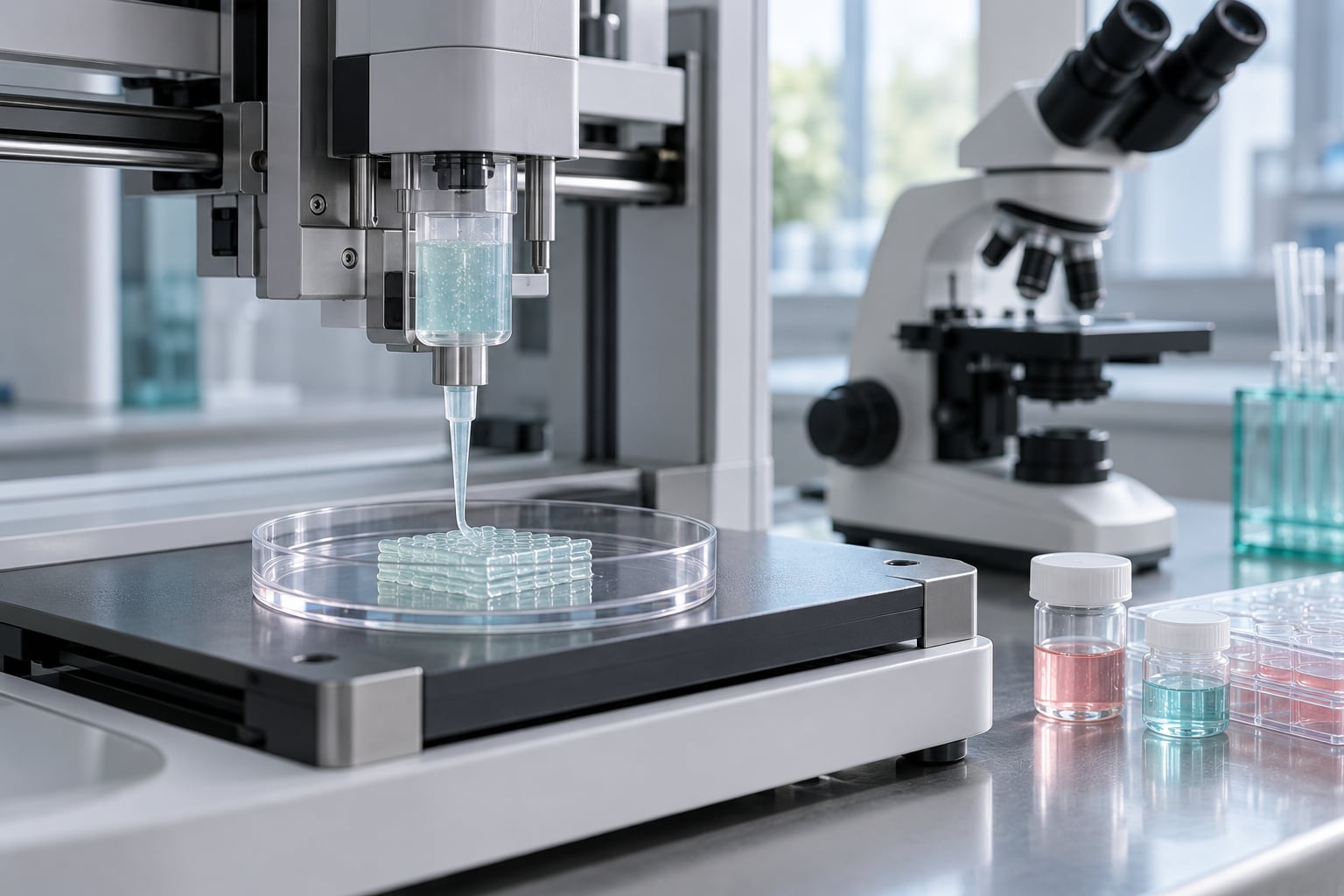
Bioprinting explained for beginners comes down to one idea: it is ordinary 3D printing, except the material being deposited contains living cells. Instead of melting plastic filament or curing resin, a printer lays down layer after layer of a cell-laden gel called a bioink, and the cells go on living and reorganising into tissue-like structures. That single swap from inert material to living material changes almost everything about how the process works, why it is slow, and why printed human organs are still a research goal rather than a hospital service.
If you have printed a part before, you already understand the geometry. What changes is the biology, the environment and the patience required. This guide walks through the whole thing in plain language: what bioprinting is, how it works step by step, what the materials are, which techniques exist, what people are actually printing today, and what still goes wrong often enough that nobody should promise you a printed kidney next year.
Key takeaways
- Bioprinting deposits living cells suspended in a printable gel, one thin layer at a time.
- The workflow has three stages: preparing cells and a model, printing, then maturing the construct in a bioreactor.
- Extrusion-based printing is the dominant method because it handles viscous bioinks and delicate cells well.
- Printing a flat, thin piece of tissue is achievable. Printing a thick, working organ with blood vessels is not.
- Most real gains today come from tissue models for research and drug testing, not from organ transplants.
Table of Contents
- 1What Is Bioprinting?
- 2How Does Bioprinting Work?
- 3Bioprinting Explained for Beginners: The Main Components
- 4Bioprinting Explained for Beginners: Bioinks You Will Hear About Most
- 5Bioprinting Techniques: Which Approach Is Used?
- 6What Are the Main Applications of Bioprinting?
- 7Why Is Bioprinting Useful?
- 8What Are the Limitations and Risks?
- 9Frequently Asked Questions
- 10Can you explain bioprinting in a simple way?
- 11How long until 3D printed organs are available?
- 12What organs can be 3D bioprinted today?
- 13Can you 3D print human skin?
- 14How much does a bio 3D printer cost?
- 15What are the basic steps of 3D printing?
- 16Where Bioprinting Is Heading
What Is Bioprinting?
Bioprinting is the layer-by-layer fabrication of structures made from living cells, usually embedded in a supportive biomaterial. A digital model tells the machine where to deposit material, and the deposited bioink solidifies just enough to hold its shape while the cells inside stay alive and begin to act like tissue.
It grew out of tissue engineering, the practice of building replacement tissue in the lab, which historically relied on moulds and scaffolds rather than a printer. Bioprinting adds direct digital control over the shape and the placement of cells, which is why a computer scan of a defect can become a construct shaped for one specific patient.
The comparison with conventional 3D printing is instructive. A plastic printer melts a solid and does not care about temperature gradients inside the part. A bioprinter moves a living suspension through a nozzle, so every force applied to that suspension matters, because excessive shear can tear cells apart. The part also cannot simply be finished and used. It has to be kept sterile, fed nutrients, given oxygen, and often grown for weeks before anyone learns whether the cells inside survived and formed anything useful.
How Does Bioprinting Work?
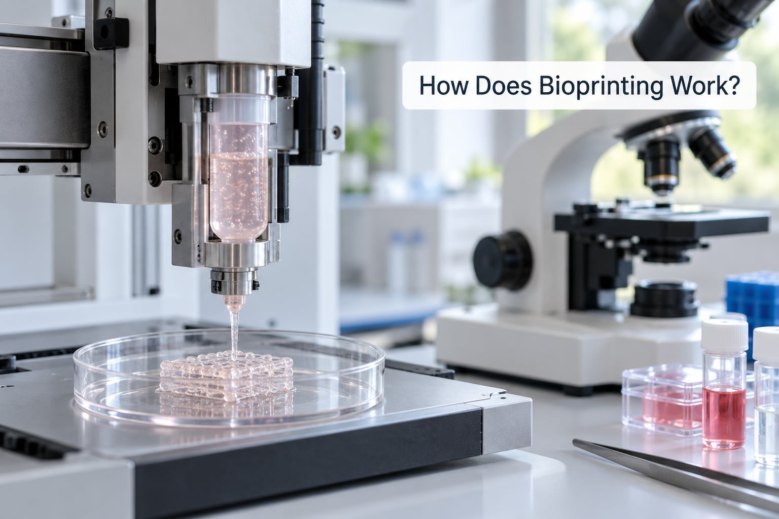
The process splits into three stages, and the first and last stages are where most of the difficulty sits. The printing itself is often the shortest part.
1. Pre-bioprinting. Researchers decide what tissue they want and where it comes from. Cells may be taken from a small biopsy, isolated from a donor sample, or drawn from a cell bank. They are expanded in culture to get enough cells, then suspended in a warm, liquid bioink just before printing. In parallel, a medical scan such as a CT or MRI is converted into a 3D model, and the model is prepared with the paths the tissue needs, including channels for nutrients in larger constructs.
2. Bioprinting. The printer deposits the cell-laden gel layer by layer, often using a heated or otherwise controlled support material underneath the fresh layers so soft structures do not collapse under their own weight. Each new layer is stiff enough to hold the last one in place, and the whole thing ends up as a wet three-dimensional gel sitting in a dish.
3. Post-bioprinting. The construct is transferred into a bioreactor, a sealed vessel that circulates nutrient-rich media, controls temperature and oxygen, and sometimes applies gentle mechanical stimulation. Over days to weeks the cells multiply, lay down their own extracellular matrix, and begin to mature into something closer to real tissue. Researchers then measure cell viability, mechanical strength and gene activity to see whether it worked.
Bioprinting Explained for Beginners: The Main Components
Five things have to line up for any bioprinting project, and beginners usually underestimate how much each one constrains the others.
Cells. These do the actual biological work. Different tissues need different cell types, and researchers often mix two populations, for instance structural cells alongside the endothelial cells that line blood vessels. Cells must survive being pushed through a nozzle and then survive weeks inside a dense gel, which is a demanding filter.
Bioink. The material carrying the cells. It has to flow through a nozzle, hold a shape once deposited, stay gentle enough for cells, and ideally break down or be remodelled by the cells themselves.
The printer. Hardware that can deposit material gently and repeatably, often with control over temperature and a sterile enclosed print chamber.
Scaffolds. Supporting structures, either printed alongside the cells or acellular and added later. Scaffolds guide tissue shape but can provoke an immune reaction, which is why scaffold-free approaches are an active area of work.
Culture conditions. Temperature, nutrient supply, oxygen level, pH and sterility, before and after printing. Most printed cells die from starvation and poor oxygen diffusion long before they fail from anything mechanical.
Bioprinting Explained for Beginners: Bioinks You Will Hear About Most
Hydrogels dominate bioinks because water-based gels mimic the wet environment cells live in. Among them, alginate gels quickly and gently and is a common teaching material, while GelMA is a photocrosslinkable gelatin derivative that cures on demand under light. Collagen and fibrin are widely used because they resemble natural structural proteins, and synthetic bioelastomers are added when mechanical strength and elasticity both matter.
Formulation is where the real engineering sits. Crosslinking is the step that turns a liquid into a solid structure, usually triggered by light, heat or a chemical reaction, and every crosslinker is a trade. Stiff enough to print means risky for cells, while soft enough to keep cells happy often means the construct slumps. Add growth factors at the wrong concentration and you get signal confusion rather than tissue, which is why the basic steps of bioprinting always end at careful tuning rather than at the printer itself.
Bioprinting Techniques: Which Approach Is Used?
Five families of method cover almost all bioprinting, and they differ mainly in how material reaches the build platform and how gently the cells survive the journey.
Extrusion-based printing pushes bioink through a nozzle using pneumatic pressure, a piston or a screw, laying down continuous filaments. It suits viscous materials and large constructs, works with cohesive spheroids as well as cell-laden gels, and coaxial nozzles place cells in a hollow core surrounded by a shell material, which is how tubular structures get built. Its limits are resolution set by nozzle diameter and shear forces at the nozzle tip.
Droplet-based and inkjet printing ejects picolitre drops on demand, giving higher resolution and precise positioning of cells. Accuracy comes at a price: cells can be damaged by the ejection pressure, and the bioinks used must stay fluid enough for tiny nozzles.
Laser-assisted bioprinting uses a focused energy source, often a pulsed laser, to move or deposit material, which removes the mechanical stress entirely and allows higher cell viability. The equipment is specialised and expensive.
Photopolymerisation methods such as stereolithography and digital light processing cure a light-sensitive resin layer by layer with excellent resolution. Photocrosslinkable bioelastomers are used here, and the constraint is that only materials which polymerise under light can be printed.
Support-bath printing, best known from FRESH printing developed by Hinton and Feinberg, injects a thermoreversible gel around the nozzle so soft bioinks can be printed freeform in three dimensions, then warmed away after printing. It is the technique that made genuinely soft, complex structures possible.
What Are the Main Applications of Bioprinting?
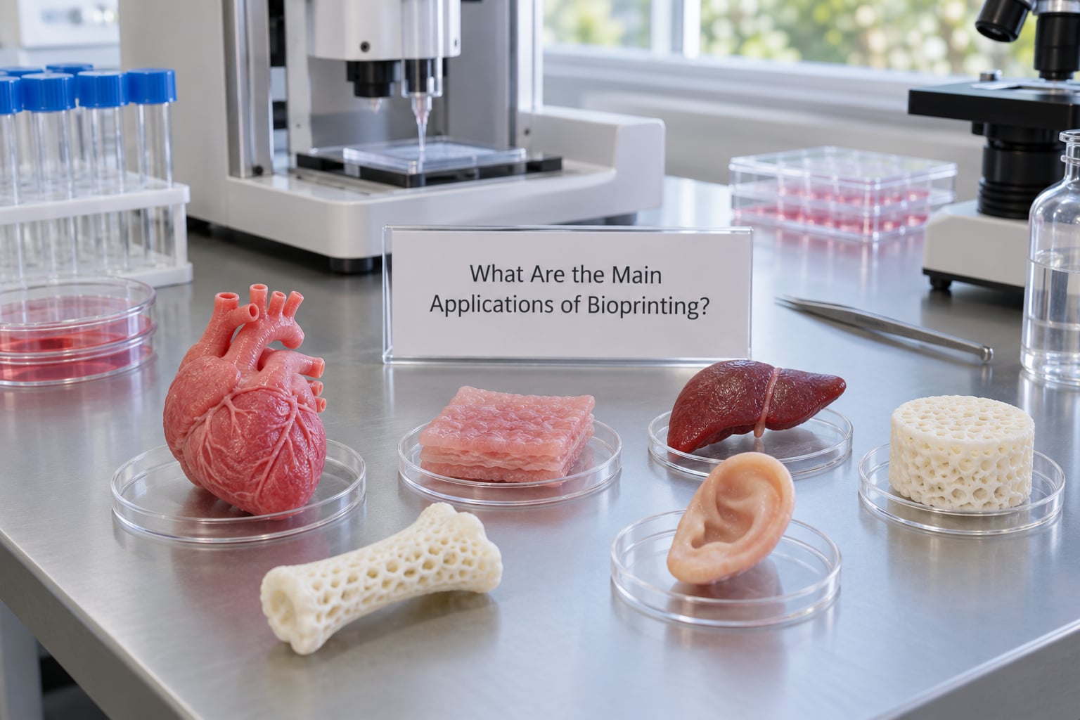
It helps to sort these into what is routine in research and what is still experimental. Very little of this is clinical care yet.
- Tissue models for research, established. Researchers build skin, cartilage, cardiac and liver models to study how cells behave, which the kinds of flat tissue models printed today handle well.
- Drug and toxicity testing, established as a research tool. Tissue models reduce reliance on animal studies for early screening, and they let researchers compare results against human cells rather than a substitute.
- Disease modelling and organ-on-a-chip, growing. Patient-derived cells can be printed into models of a specific condition, giving a personalised picture of how a disease progresses.
- Scaffolds and acellular implants, in trials. These are printed structures without living cells, so they face none of the biology problems, and several have progressed toward clinical use.
- Implants and custom devices, practical. Patient-specific surgical guides, prosthetic parts and anatomical models printed from a scan are already useful, though strictly speaking they contain no cells.
- Skin grafts and cartilage repair, in human trials. Bioprinted skin for wound coverage is among the closest tissue-engineering products to real patients.
- Regenerative medicine, experimental. Printing replacement tissue that integrates with a patient and functions long term remains early-stage.
- Food, an active research area. Work at Kyushu University and Cardiff University has produced printed foods with textures tuned for people who have difficulty swallowing, using controlled radio-frequency and microwave heating to change firmness and water retention.
- Cultured meat, prototype stage. Cultured beef and Wagyu have been demonstrated, but cost and texture keep it far from shelves.
- Environmental uses, early. Printing living bacteria to break down contaminants in wastewater, or arranging microbial communities as biofilms, is being explored.
Why Is Bioprinting Useful?
Donor organs are chronically scarce, and the need grows while the supply of suitable donors does not. Printing tissue instead of waiting for a donor is the long-term argument for the field, and it is why regenerative medicine funding keeps bioprinting on its list.
The nearer payoff is in research. Human tissue models can give a drug candidate a fast, human-specific read on toxicity and efficacy before it reaches clinical trials, and they can be produced on demand rather than ordered. In some testing contexts this reduces the number of animals needed, which matters both ethically and practically, since animal models disagree with human biology more often than researchers would like.
Customization is the other quiet advantage. Because the construct comes from a digital model, a defect or an organ can be shaped to one patient’s scan, and laboratories can print variations of a design to test which one works. Reading how a printed tissue behaves also teaches researchers something no plastic sample can tell them about cell behaviour.
What Are the Limitations and Risks?
The honest list is long, and this is the section most beginner explainers skip.
Vascularization is the core problem. Oxygen and nutrients move by diffusion, which only reaches about a few hundred micrometres from a capillary. Printing a thick block without a blood supply starves the interior cells, so researchers print channels, print vessel cells, or build mini-tissue blocks that stay small enough to feed themselves.
Cell viability varies. Every stage can cost cells: nozzle shear, UV exposure during photocrosslinking, pH shifts, the transfer itself. Printing is not the last step, it is the start of a long period where cells may still fail.
Mechanical properties lag. Printed constructs are usually softer than native tissue, and native tissue has a specific gradient of stiffness, alignment and porosity that is hard to reproduce.
Scale-up and repeatability are unsolved. A working small print does not automatically scale, and moving from a validated laboratory protocol to a production line introduces its own problems.
Sterilization, storage and shelf life are rarely discussed and all commercially difficult. A living product cannot sit on a warehouse shelf, so manufacturing, transport and regulation all assume a very different product lifecycle.
Regulation is real but slow. A printed organ combines a device, a biologic and a surgical procedure, so it faces approval requirements none of its parts would face alone.
Cost and skills. Real bioprinting needs a sterile lab, cell culture capability, a bioreactor and trained staff. Prices vary widely with hardware and setup, and the total for a working tissue-culture line sits well above a desktop printer, which is why the honest answer to the cost question starts with what you are trying to print.
Hype-versus-reality gap. No, you cannot print a working human organ yet, and nobody has. No whole human has been 3D printed, and printed tissues in people are still experimental. The 2006 engineered bladders from Wake Forest, the 2015 FRESH work and the first 3D-bioprinted ear transplant in 2022 are genuine milestones, but each is a step rather than an arrival. Expect organ-level printing to remain research news for years.
Frequently Asked Questions
Can you explain bioprinting in a simple way?
Think of it as 3D printing with living cells instead of plastic. A printer deposits a gel containing cells one thin layer at a time, and the cells stay alive inside it. The printed gel then sits in a bioreactor, where the cells multiply and start behaving like real tissue. Researchers use it to build tissue models for research and, eventually, replacement tissue for patients.
How long until 3D printed organs are available?
A working human organ with its own blood supply is still years away, and no printed organ has been implanted in a person. The obstacle is vascularization: oxygen diffuses only a few hundred micrometres, so any thick construct needs blood vessels that researchers cannot yet print reliably. Thin tissues and scaffolds are much closer, with some already in human trials.
What organs can be 3D bioprinted today?
In practice, teams print tissue models and simple constructs: skin, cartilage, cardiac and liver tissue, and flat or tubular layers used for drug testing. Whole organs have not been produced. Bioprinted bladders were implanted in patients in the 2006 Wake Forest work, and a 3D-bioprinted ear was transplanted in 2022, but both were limited structures rather than functioning organs.
Can you 3D print human skin?
Yes, in the sense that skin models and skin grafts can be printed from cell-laden hydrogels, and bioprinted skin for wound coverage has reached human trials. The printed sheet is usually a thin, flat construct a few cell layers thick, because thicker tissue needs a blood supply it does not have yet. For most everyday injuries, conventional skin grafts remain the standard treatment.
How much does a bio 3D printer cost?
Prices range widely because bioprinters come in very different forms. Desktop systems intended for teaching with cell-free or simple gels cost far less than research platforms with sterile chambers and temperature control, and a functioning tissue-culture laboratory costs more than the printer alone. The number that matters most is what you plan to print, since live-cell work adds cell culture, biosafety and bioreactor costs on top.
What are the basic steps of 3D printing?
For bioprinting there are three stages. First, pre-bioprinting: cells are isolated and multiplied, mixed into a bioink, and a scan or design is turned into a 3D model. Second, printing, where the material is deposited layer by layer and each layer is stabilised. Third, post-bioprinting, where the construct goes into a bioreactor to grow and mature before it is tested.
Where Bioprinting Is Heading
If you take one thing away, make it this: bioprinting is already a real tool for studying tissue and testing drugs, and it is still early-stage as a way of replacing organs. The honest version of the story is more useful than the hype version, because the parts that work are quietly changing research labs now.
Start with the three-stage workflow and the bioink problem, since everything else follows from them. Watch printed tissue models for disease and organ-on-a-chip systems, which are closest to routine use. And treat any headline promising a printed organ as something to check against the vascularization problem before you get excited.